
A 45-year-old man undergoes abdominal CT scan after a motor vehicle collision. CT scan reveals only an incidental 3.5-cm right adrenal lesion (<10 Hounsfield units). The patient is otherwise healthy and has no other injuries related to the collision. His electrolytes are normal. Plasma-free metanephrines are normal
Which of the following steps is
also appropriate to evaluate his adrenal lesion?
A. MRI.
B. dexamethasone suppression test.
C. metaiodobenzylguanidine (MIBG) scan.
D. plasma aldosterone concentration and renin activity.
E. observation.
ANSWER: B
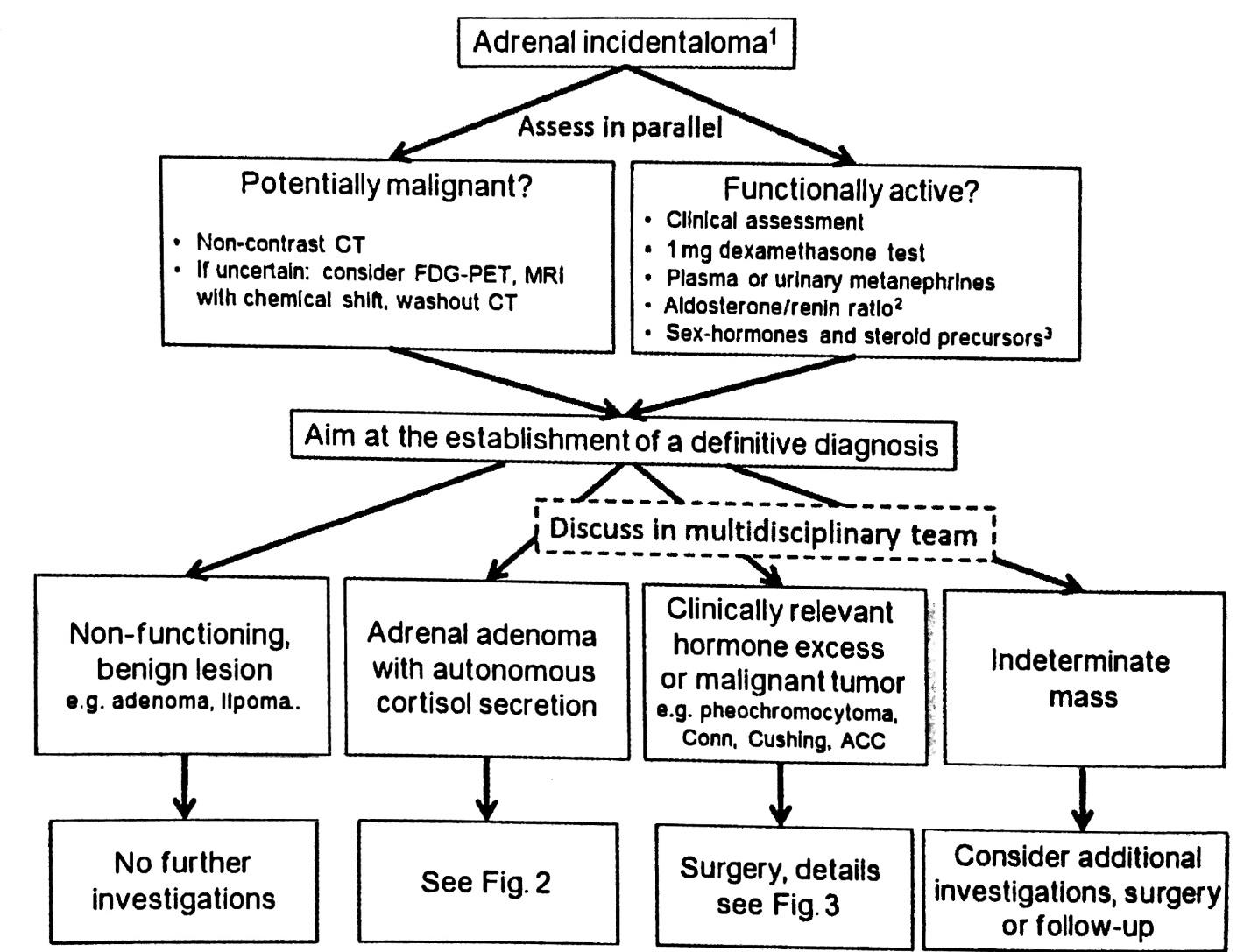
Adrenal incidentaloma is encountered in 3 to 8% of patients undergoing imaging for other reasons. Evaluation is directed at determining malignant potential and the lesion's functional activity
Metaiodobenzylguanidine (MIBG) scanning was formerly used for localization of pheochromocytoma but has been replaced by MRI; PET scan is used in selected cases.
Radiographic characteristics assist in determining malignant potential.
Size and Hounsfield units (HU) on CT scan are
significant predictors of malignant potential. Size less than 4 cm with 10 HU or fewer are unlikely to be malignant; therefore, further imaging such as MRI is not needed.
Functional activity should be evaluated through careful history and physical examination. All patients with adrenal incidentaloma should undergo screening for cortisol excess using a dexamethasone suppression test.
All patients should be screened for pheochromocytoma using plasma-free
metanephrines or urinary fractionated metanephrines.
Screening for aldosteronoma using plasma aldosterone concentration and renin activity can be reserved for patients with hypertension and hypokalemia
Which of the following steps is
also appropriate to evaluate his adrenal lesion?
A. MRI.
B. dexamethasone suppression test.
C. metaiodobenzylguanidine (MIBG) scan.
D. plasma aldosterone concentration and renin activity.
E. observation.
ANSWER: B
Adrenal incidentaloma is encountered in 3 to 8% of patients undergoing imaging for other reasons. Evaluation is directed at determining malignant potential and the lesion's functional activity
Metaiodobenzylguanidine (MIBG) scanning was formerly used for localization of pheochromocytoma but has been replaced by MRI; PET scan is used in selected cases.
Radiographic characteristics assist in determining malignant potential.
Size and Hounsfield units (HU) on CT scan are
significant predictors of malignant potential. Size less than 4 cm with 10 HU or fewer are unlikely to be malignant; therefore, further imaging such as MRI is not needed.
Functional activity should be evaluated through careful history and physical examination. All patients with adrenal incidentaloma should undergo screening for cortisol excess using a dexamethasone suppression test.
All patients should be screened for pheochromocytoma using plasma-free
metanephrines or urinary fractionated metanephrines.
Screening for aldosteronoma using plasma aldosterone concentration and renin activity can be reserved for patients with hypertension and hypokalemia
A 45-year-old man undergoes abdominal CT scan after a motor vehicle collision. CT scan reveals only an incidental 3.5-cm right adrenal lesion (<10 Hounsfield units). The patient is otherwise healthy and has no other injuries related to the collision. His electrolytes are normal. Plasma-free metanephrines are normal
Which of the following steps is
also appropriate to evaluate his adrenal lesion?
A. MRI.
B. dexamethasone suppression test.
C. metaiodobenzylguanidine (MIBG) scan.
D. plasma aldosterone concentration and renin activity.
E. observation.
ANSWER: B
Adrenal incidentaloma is encountered in 3 to 8% of patients undergoing imaging for other reasons. Evaluation is directed at determining malignant potential and the lesion's functional activity
Metaiodobenzylguanidine (MIBG) scanning was formerly used for localization of pheochromocytoma but has been replaced by MRI; PET scan is used in selected cases.
Radiographic characteristics assist in determining malignant potential.
Size and Hounsfield units (HU) on CT scan are
significant predictors of malignant potential. Size less than 4 cm with 10 HU or fewer are unlikely to be malignant; therefore, further imaging such as MRI is not needed.
Functional activity should be evaluated through careful history and physical examination. All patients with adrenal incidentaloma should undergo screening for cortisol excess using a dexamethasone suppression test.
All patients should be screened for pheochromocytoma using plasma-free
metanephrines or urinary fractionated metanephrines.
Screening for aldosteronoma using plasma aldosterone concentration and renin activity can be reserved for patients with hypertension and hypokalemia
0 التعليقات
0 نشر
16191 ظهور